Home
/ Diagram Of Human Backbone : Lumbar Spine Anatomy Diagram High Res Stock Images Shutterstock : It runs down the centre of the body.
Diagram Of Human Backbone : Lumbar Spine Anatomy Diagram High Res Stock Images Shutterstock : It runs down the centre of the body.
Diagram Of Human Backbone : Lumbar Spine Anatomy Diagram High Res Stock Images Shutterstock : It runs down the centre of the body.. Between the vertebrae are small spaces known as intervertebral canals that allow spinal nerves to exit the spinal cord and connect to the various regions of the body. Certain back muscles extend to other areas, like the shoulders, upper arms, and thighs. It consists of various groups of vertebrae. The spinous processes are horizontal and more squared in shape. The vertebral column, also known as the spinal column, is a flexible column that encloses the spinal cord and also supports the head.
Certain back muscles extend to other areas, like the shoulders, upper arms, and thighs. The spine diagram the spine diagram shown below, consists of many bones or vertebrae,soft discs,the spinal cord, and spinal nerves. Just need a glimpse, leave your valuable advice let us know , and subscribe us! The spinous processes are horizontal and more squared in shape. Our latest youtube film is ready to run.
Diagram Database Just The Best Diagram Database Website from i0.wp.com The thoracic spine helps keep the body upright and stable. This human anatomy module is composed of diagrams, illustrations and 3d views of the back, cervical, thoracic and lumbar spinal areas as well as the various vertebrae. Shows the medical accuracy of human skeleton and 3d rendering. The human back extends from the buttocks to the posterior portion of the neck and shoulders. Related posts of human back bones diagram long bone diagram labeled colored. As you can see in the vertebrae diagram above, the human spine consists of 33 vertebrae in total; When you look at the skeleton from behind, you can clearly see the spine running down the back, and the broad plates of the shoulder blades and pelvis. It is particularly interesting for physiotherapists, osteopaths, rheumatologists, neurosurgeons.
Certain back muscles extend to other areas, like the shoulders, upper arms, and thighs.
The vertebrae of the spine align so that their vertebral canals form a hollow, bony tube to protect the spinal cord from external damage and infection. Just need a glimpse, leave your valuable advice let us know , and subscribe us! Diagram of a human spine with the. 24 are considered to be part of the upper spine, whilst the other 11 are found in the sacrum & coccyx. We hope this picture the human spine anatomy in detail can help you study and research. This human anatomy module is composed of diagrams, illustrations and 3d views of the back, cervical, thoracic and lumbar spinal areas as well as the various vertebrae. Some of these muscles are quite large and cover broad areas. Spinal vertebrae bone spine vertebra toracica spinal cord spine structure back diagram spine sections spinal cord vertebrae spinal structure health diagram. The vertebral column, also known as the backbone or spine, is part of the axial skeleton.the vertebral column is the defining characteristic of a vertebrate in which the notochord (a flexible rod of uniform composition) found in all chordates has been replaced by a segmented series of bone: The spine anatomy is a complex structure. Your spine, or backbone, is your body's central support structure. Diagram of a human spine with the name and description of all sections and segments of the vertebrae, vector illustration. The vertebral column houses the spinal canal, a cavity that.
The vertebral column, also known as the backbone or spine, is part of the axial skeleton.the vertebral column is the defining characteristic of a vertebrate in which the notochord (a flexible rod of uniform composition) found in all chordates has been replaced by a segmented series of bone: We think this is the most useful anatomy picture that you need. The spinous processes are horizontal and more squared in shape. The human spine consists of 33 vertebrae: The vertebrae, which stack like spools of thread, support the back and protect the spinal cord.
Vertebral Column An Overview Sciencedirect Topics from ars.els-cdn.com Related posts of human back bones diagram long bone diagram labeled colored. The vertebral column is the most important collection of bones to maintain stability of the skeletal structure and support of the entire body, especially when upright. Anatomynote.com found the human spine anatomy in detail from plenty of anatomical pictures on the internet. Diagram of a human spine with the name and description of all sections and segments of the vertebrae, vector illustration. Vertebrae separated by intervertebral discs. 3d rendered medically accurate illustration of the human spine. The spinous processes are horizontal and more squared in shape. The red lines point individual bones and the names are writen in singular, the blue lines conect to group of bones and are in plural form.
Shows the medical accuracy of human skeleton and 3d rendering.
Anatomynote.com found the human spine anatomy in detail from plenty of anatomical pictures on the internet. The spine has three normal curves: Between the vertebrae are small spaces known as intervertebral canals that allow spinal nerves to exit the spinal cord and connect to the various regions of the body. The muscles of the lower back help stabilize, rotate, flex, and extend the spinal column, which is a bony tower of 24 vertebrae that gives the body structure and houses the spinal cord. Vertebrae separated by intervertebral discs. The atlas is the topmost vertebra, and along with c2, forms the joint connecting the skull and spine. Diagram of a human spine with the name and description of all sections and segments of the vertebrae, vector illustration. The vertebral column, also known as the backbone or spine, is part of the axial skeleton.the vertebral column is the defining characteristic of a vertebrate in which the notochord (a flexible rod of uniform composition) found in all chordates has been replaced by a segmented series of bone: The spinous processes are horizontal and more squared in shape. It runs down the centre of the body. Lateral labeled diagram of the human vertebral spinal column showing vertebrae numbering order and the 5 different regions of the spine. For more anatomy content please follow us and visit our website: Related posts of human back bones diagram long bone diagram labeled colored.
The vertebral column is the most important collection of bones to maintain stability of the skeletal structure and support of the entire body, especially when upright. Your spine, or backbone, is your body's central support structure. Just need a glimpse, leave your valuable advice let us know , and subscribe us! Browse 748 spine diagram stock photos and images available, or search for back pain or chiropractor to find more great stock photos and pictures. Person outline body outline body diagram body chart human body drawing body reference drawing body template free printable calendar templates line sketch.
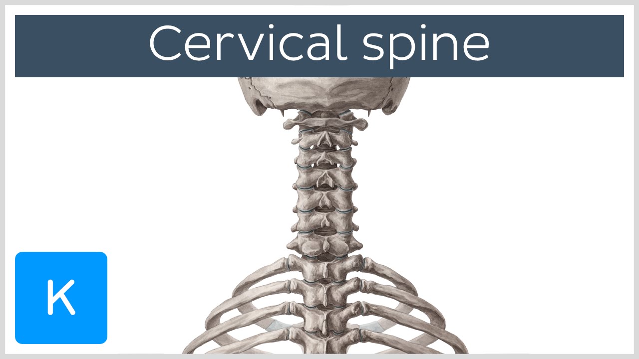
Cervical Vertebrae Physiopedia from i.ytimg.com The vertebrae, which stack like spools of thread, support the back and protect the spinal cord. The atlas is the topmost vertebra, and along with c2, forms the joint connecting the skull and spine. Your skeleton can be divided into two main parts. When you look at the skeleton from behind, you can clearly see the spine running down the back, and the broad plates of the shoulder blades and pelvis. We are pleased to provide you with the picture named anatomy of back muscles diagram.we hope this picture anatomy of back muscles diagram can help you study and research. Spinal vertebrae bone spine vertebra toracica spinal cord spine structure back diagram spine sections spinal cord vertebrae spinal structure health diagram. Diagram of a human spine with the. Long bone diagram labeled colored 12 photos of the long bone diagram labeled colored , bone
Diagram of a human spine with the.
We hope this picture the human spine anatomy in detail can help you study and research. The vertebral column of the lower back includes the five lumbar vertebrae, the sacrum, and the coccyx. The back's muscles start at the top of the back (named the cervical vertebrae) and go to the tailbone (also named the coccyx). It consists of various groups of vertebrae. See human back anatomy stock video clips. This human anatomy module is composed of diagrams, illustrations and 3d views of the back, cervical, thoracic and lumbar spinal areas as well as the various vertebrae. The atlas is the topmost vertebra, and along with c2, forms the joint connecting the skull and spine. The vertebrae of the spine align so that their vertebral canals form a hollow, bony tube to protect the spinal cord from external damage and infection. The spine diagram the spine diagram shown below, consists of many bones or vertebrae,soft discs,the spinal cord, and spinal nerves. Diagram of a human spine with the name and description of all sections and segments of the vertebrae, vector illustration. Shows the medical accuracy of human skeleton and 3d rendering. See lumbar spine anatomy diagram stock video clips. The human spine consists of 33 vertebrae:
We think this is the most useful anatomy picture that you need diagram of backbone. Certain back muscles extend to other areas, like the shoulders, upper arms, and thighs.